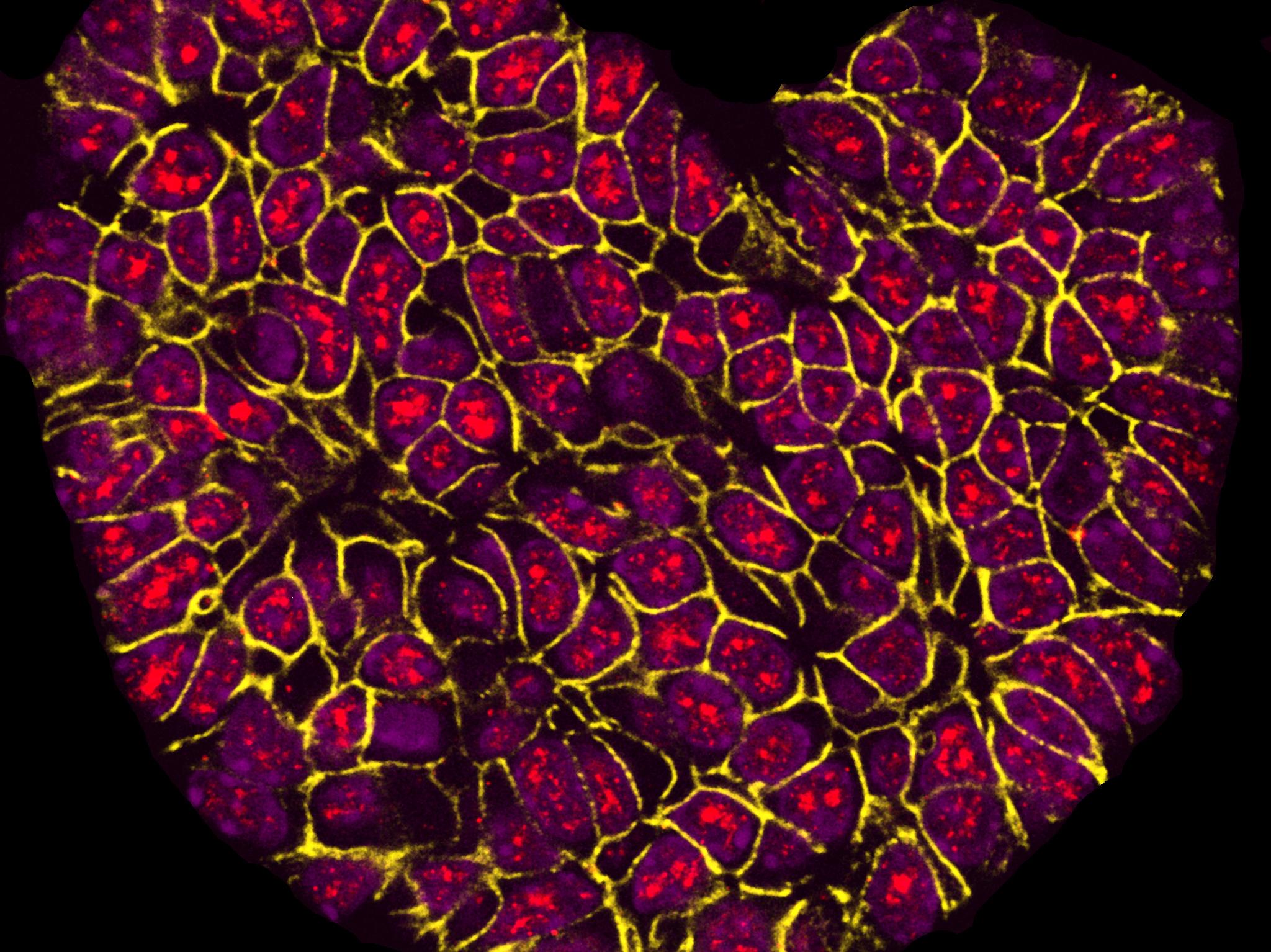
Picture of heart-shaped cancer cells wins science photography prize
‘Fluorescent labelling and microscopy can reveal stunning shapes and colours in things like human cells’

Your support helps us to tell the story
From reproductive rights to climate change to Big Tech, The Independent is on the ground when the story is developing. Whether it's investigating the financials of Elon Musk's pro-Trump PAC or producing our latest documentary, 'The A Word', which shines a light on the American women fighting for reproductive rights, we know how important it is to parse out the facts from the messaging.
At such a critical moment in US history, we need reporters on the ground. Your donation allows us to keep sending journalists to speak to both sides of the story.
The Independent is trusted by Americans across the entire political spectrum. And unlike many other quality news outlets, we choose not to lock Americans out of our reporting and analysis with paywalls. We believe quality journalism should be available to everyone, paid for by those who can afford it.
Your support makes all the difference.A photograph entitled I Heart Research – showing cancerous cells – has won a new science photography prize.
The BMC Research in Progress Photo Competition received submissions from around the world, such as Australia, Nepal, Iraq, China and Brazil.
The entries included images of flowers, birds, butterflies, a bear living in captivity, and diseases like Zika and Dengue being studied in a lab.
The winner was a photograph of a tumour from a mouse in the shape of a heart that had been fluorescently labelled for inspection through a microscope.
The photographer Sarah Boyle, of the Centre for Cancer Biology in Adelaide, Australia, said: “I took it as part of my research into breast cancer and for me it really shows how processes that we researchers use almost on a daily basis – such as fluorescent labelling and microscopy – can reveal stunning shapes and colours in things like human cells.
“As part of our research into breast cancer, we are able to use specialist microscopes and imaging equipment to make visible different proteins.
“The tumour we see in this image is fluorescently labelled red for the active form of a protein whose levels may increase as cancer develops.”
The competition was run by BioMed Central (BMC), which publishes scientific journals.
Rachel Burley, BMC’s publishing director, said: “This image demonstrates the ability of science and research to discover surprises even in the most unlikely of places, as well as the enthusiasm researchers have for their work.
“It is visually stunning and its bold colours and jewel-like shape really made it stand out to our judges.
“Importantly, it is also an example of how vital it is for research to keep progressing and for researchers to have access to the latest results, so that they can build on existing findings, advancing discovery in fields like cancer research.”
The runner-up was a picture taken by Yuan Xiao Wei, a post-doctoral researcher at the China Agricultural University, entitled The Power of Life, showing cucumber seeds growing in a petri dish.
“The purpose of the experiment is to test how seeds germinate and how fast and well seedlings are likely to grow and establish crops. The results are important for cucumber breeding,” a statement from BMC said.
The winner was chosen from 266 entries across four research-related categories: people at work; close-ups of equipment; plants and animals; and microscopy.
Join our commenting forum
Join thought-provoking conversations, follow other Independent readers and see their replies
Comments