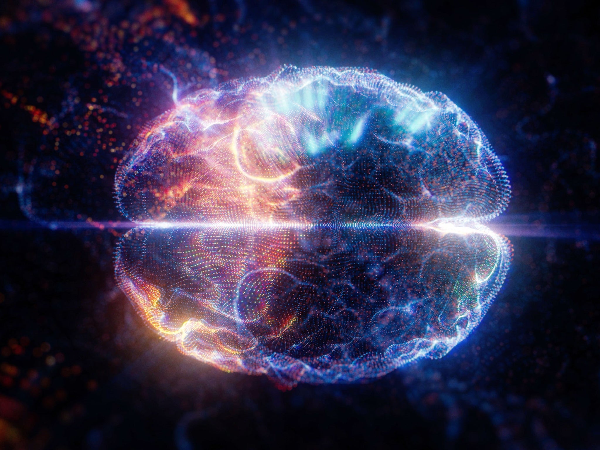
Scanner that detects light inside the brain could lead to Alzheimer’s screening and new cancer treatment
Light microscopes have been peering inside living cells and thin slices of tissue since the late 1500s, but have not been able to reach further until now

Sign up for our free Health Check email to receive exclusive analysis on the week in health
Get our free Health Check email
A new scanner that detects light deep inside the brain could lead to revolutionary cancer treatment and screening programmes for Alzheimer’s disease.
The device uses magnetic resonance imaging (MRI) to map how light spreads in opaque environments, capturing dynamic changes in colours of tissue.
It could map neuron-stimulating fibres during experiments or monitor patients receiving light-based therapies for tumours.
Senior author Professor Alan Jasanoff, of Massachusetts Institute of Technology (MIT) in the US, said: “We can image the distribution of light in tissue. That’s important.
“People who use light to stimulate or measure tissue often don’t quite know where the light is going, where they’re stimulating or where the light is coming from. Our tool can be used to address those unknowns.”
Light microscopes have been peering inside living cells and thin slices of tissue since the late 1500s, but have not been able to reach further until now.
Professor Jasanoff explained: “One of the persistent problems in using light, especially in the life sciences, is it doesn’t do a very good job penetrating many materials.
“Biological materials absorb light and scatter light, and the combination of those things prevents us from using most types of optical imaging for anything that involves focusing in deep tissue.”
So his students helped design a sensor that could transform light into a magnetic signal.
Professor Jasanoff said: “We wanted to create a magnetic sensor that responds to light locally, and therefore is not subject to absorbance or scattering. Then this light detector can be imaged using MRI.”
The lab has previously developed MRI probes that can interact with a variety of molecules in the brain, including feelgood chemical dopamine and calcium.
When they bind to their targets, it affects magnetic interactions with the surrounding tissue, dimming or brightening the signal.
The light-sensitive version encases magnetic particles in a tiny bubbles of fat called liposomes. They become ‘leaky’ when exposed to certain wavelengths.
The magnetic particles called liposomal nanoparticle reporters (LisNR) interact with water - and generate a signal detectable by MRI.
The researchers created particles that became leaky when exposed to ultraviolet light - and impermeable in blue light. There were also responces to other wavelengths.
Experiments on rats targeted a part of the brain called the striatum - involved in planning movement and responding to reward.
After injecting the particles the researchers were able to map the distribution of light from an optical fibre implanted nearby.
It is similar to those used for optogenetic stimulation. So this kind of sensing could be useful to researchers who perform optogenetic experiments in the brain.
Professor Jasanoff said: “We don’t expect that everybody doing optogenetics will use this for every experiment.
“It’s more something you would do once in a while to see whether a paradigm you’re using is really producing the profile of light you think it should.”
The device described in Nature Biomedical Engineering could also be useful for monitoring patients receiving treatments that involve light.
Professor Jasanoff said photodynamic therapy for instance uses light from a laser or LED to kill cancer cells.
Dr Xin Yu, of Harvard Medical School, Boston, who was not involved in the study, added: “This paper shows a novel sensor to enable photon detection with MRI through the brain.
“This illuminating work introduces a new avenue to bridge photon and proton-driven neuroimaging studies.”
The researchers are now working on similar probes that could be used to detect light emitted by luciferases - glowing proteins used in biological tests.
It could reveal whether a particular gene is activated or not. Currently they can only be imaged in superficial tissue or cells grown in a lab dish.
Professor Jasanoff also hopes to use the strategy to design MRI probes that can detect stimuli other than light, such as neurochemicals or other molecules found in the brain.
He said: “The principle we use to construct these sensors is quite broad and can be used for other purposes too.” The device is described in Nature Biomedical Engineering.
Subscribe to Independent Premium to bookmark this article
Want to bookmark your favourite articles and stories to read or reference later? Start your Independent Premium subscription today.
Join our commenting forum
Join thought-provoking conversations, follow other Independent readers and see their replies